

Mastodon Tusk Project Posters
In the fall of 2016, the University of Northern Iowa Museum was awarded a $306,258 heritage grant from the Roy J. Carver Charitable Trust for the "Scientific Study, Conservation, and Interpretation" of a mastodon tusk held by the museum. The UNI Museum has established a partnership with the Department of Chemistry and Biochemistry led by Assistant Professor Joshua A. Sebree. During his upper level undergraduate fall CHEM 4310 Instrumental Analysis class, he plans on using samples of the Hampton mastodon tusk to run elemental composition tests. The class was designed as a way for students to understand how analytical instruments work, the proper way to use instruments, and how to perform independent research while collecting publishable results. Over the course of the following three years, the tusk will be cleaned, analyzed, and preserved along with records of the process for eventual permanent display at UNI. To do so the chemical nature of the tusk must be known in as much detail as possible.
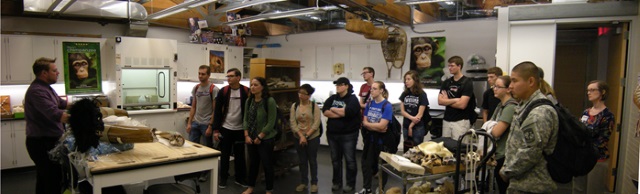
Students worked in groups to prepare and submit posters to display their work on the Hampton mastodon tusk.
-
 Mapping of Lead in Mastodon Tusk Using SEM by Nicholas Bonde and Katherine Plotzke")
Energy-Dispersive X-ray (EDX) Mapping of Lead in Mastodon Tusk Using SEM
Nicholas Bonde and Katherine Plotzke
The composition and location of lead within a prehistoric mastodon tusk was mapped using Energy Dispersive X-ray (EDX) spectroscopy analysis via a Scanning Electron Microscope (SEM). This analysis was intended to provide information on the distribution of lead throughout the tusk. Data collected from this analysis has provided insights into the sources of the lead.
-

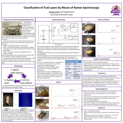
Classification of Tusk Layers by Means of Raman Spectroscopy
Treasure Divis and Vaughn Koch
The American Mastodon tusk was donated to the University of Northern Iowa, and Dr. J. Cable took the lead on preserving it for museum display at UNI. Dr. Cable’s notes stated that it would be possible to preserve the entire tusk, because the outside was “covered with a hard enamel-like substance”. Some notes were taken as to what materials were used to preserve the tusk early on, but their exact composition was not noted.
• Glazed and varnished between 1933 and 1960
• 1960 – canned patching plaster on chipping areas, glazing liquid (brown), two coats of varnish (satin finish with some gloss added)
• 1966 – wood brace and steel band added near tip to prevent cracking
No notes were taken on the preservation process after 1966, so the materials used are completely unknown. The goal of this project is to analyze the composition of the restoration materials used on the tusk and to strip the restoration materials off of the tusk without harming the tusk itself.
-

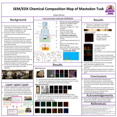
SEM/EDX Chemical Composition Map of Mastodon Tusk
Juliana Herran
The goal of this project was to study one of the rarest objects in the collection of the UNI museum dating to the Aftonian or Yarmouth age (around 120,000 to 200,000 years old).
The tusk is 11 feet, 7.5 inches with a circumference at the proximal end of 2 feet, 2 inches.
There have been previous attempts of restoration of this tusk but there are not specific procedures on record that specify the materials or methods used during the restoration attempts with sufficient detail.
With the support of the Carver Grant awarded to the UNI museum, research teams containing students from the chemistry, geology, history, art and anthropology departments will contribute to the study and preparation for the final restoration of the tusk.
The first phase of the project consists in the determination of the materials used to in the past restorations attempts. There are a variety of subprojects using different instruments and analytical techniques to achieve this goal.
For this part of the project, a chemical map of the tusk was generated using the Evex Mini SEM/EDX.
-

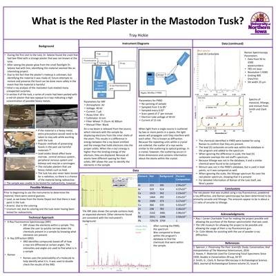
What is the Red Plaster in the Mastodon Tusk?
Tray Hickie
During the first visit to the tusk, Dr. Sebree found the crack that had been filled with a strange plaster that was not known at the time.
After seeing the plaster glow from the small flashlight Dr. Sebree had with him, identifying this material seemed like an interesting project.
Due to the fact that the plaster’s makeup is unknown, but identifying the material it was made of, future attempts to restore and preserve the fossil can be done more safely in the event that the material is harmful
Initial x-ray analysis of the mastodon tusk revealed many unexpected surprises.
In section 4 of the tusk, a series of cracks had been packed with a red-ish plaster that was opaque to x-rays indicating a high concentration of possible heavy metals.
If the material is a heavy metal, extra precautions would need to be taken to stay safe while working with the tusk.
• Popular methods of preserving fossils in the past use harmful materials.[2]
• Lead oxides blood affect bone marrow , central nervous system , peripheral nervous system and kidneys and may be a carcinogen.
• This includes plasters that use heavy metals such as lead.
• The tusk has also never been tested for a radiation, so there is a chance of the material being radioactive.
The sample was unable to be tested for radioactivity, however.
-

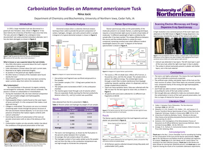
Carbonization Studies on Mammut americanum Tusk
Nina Jocic
Many preservation attempts have been made on the tusk that the University of Northern Iowa acquired. Due to the poor preservation attempts a lot of damage has happened and has caused the tusk to be in a way of disrepair. The Carver grant is very keen on the future preservation of the tusk. The sublimation of nucleobases will be able to help guide the type of preservation methods used in order to preserve the nucleobases and possible DNA still in the tusk. The extraction of nucleobases will not only help with the future preservation of tusk but it will also help to provide a potential family tree of the Mammut americanum founded in the area.
-

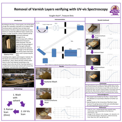
Removal of Varnish Layers verifying with UV-vis Spectroscopy
Vaughn Koch and Treasure Divis
According to the Roy J. Carver grant, one of the goals is to get the mastodon tusk back to its original state. The preservation methods have not been well documented, therefore determining what the best solvents to remove the different layers needs to be known. In order to do this, various solvents will be used and confirmed through UV-vis spectroscopy with a Diffuse Reflectance Accessory (DRA) attached. This will measure the percentage of light that gets reflected back towards the detector. The image to the left shows the various layers that have been used in preserving the tusk, including some organic and inorganic material. As the layers are removed the composition will be checked by T. Divis, which will also confirm when there is only Tusk material remaining on the sample. Due to the size of the Tusk (see below), it is important to know what solvent will remove the most layers, and work the most efficiently.
-

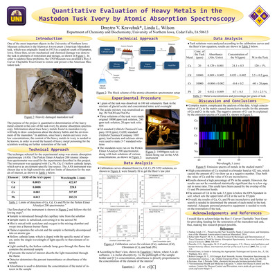
Quantitative Evaluation of Heavy Metals in the Mastodon Tusk Ivory by Atomic Absorption Spectroscopy
Dmytro V. Kravchuck and Linda L. Wilson
One of the most important objects in the University of Northern Iowa Museum collection is the Mammut Americanum (American Mastodon) tusk, which was originally found in 1933 in a sand pit south of Hampton, Iowa. Since then, severe mechanical and chemical damage was done to the tusk in attempts of restoration and storage, as shown in Figure 1. In order to address these problems, the UNI Museum was awarded a Roy J. Carver Charitable Trust Grant to restore and preserve the American Mastodon tusk. The purpose of the project is quantitative determination of the heavy metal content in the core of the tusk ivory by atomic absorption spectroscopy. Information about trace heavy metals found in mastodon ivory, will help to draw conclusions about the dietary habits and the environment of the mastodon. In addition, since heavy metals are toxic even low concentrations, the content of the heavy metals in ivory is needed to be known, in order to avoid the hazard of heavy metal poisoning for the scientists working on further restoration of the tusk.
-

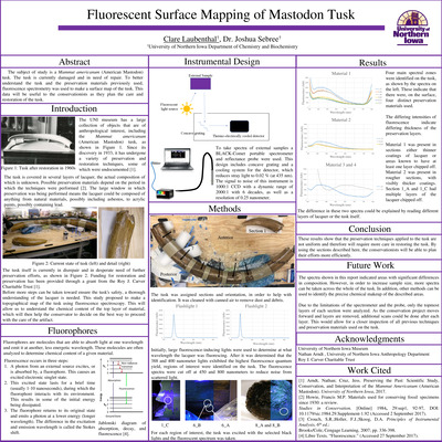
Fluorescent Surface Mapping of Mastodon Tusk
Clare Laubenthal and Joshua Sebree
The subject of study is a Mammut americanum (American Mastodon) tusk. The tusk is currently damaged and in need of repair. To better understand the tusk and the preservation materials previously used, fluorescence spectrometry was used to make a surface map of the tusk. This data will be useful to the conservationists as they plan the care and restoration of the tusk.
-

An In Between Study: The usage of cross-sectional SEM and Raman Spectroscopy to map the surface interfacing of a mastodon tusk and its lacquers
Katherine Plotzke
A microscopy study utilizing a DXR2 SmartRaman and Evex Mini-Scanning Electron Microscope (SEM) was done on the UNI Museum’s Mammut americanum (American Mastodon) tusk. A map of the surface interfacing was generated and provided information about the elemental composition and layer interconnectedness. Additionally, this project will also aid in the future restoration work on the tusk funded by the Roy J. Carver Charitable Trust.
-

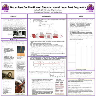
Nucleobase Sublimation on Mammut americanum Tusk Fragments
Joshua Prybil
Many preservation attempts have been made on the tusk that the University of Northern Iowa acquired. Due to the poor preservation attempts a lot of damage has happened and has caused the tusk to be in a way of disrepair. The Carver grant is very keen on the future preservation of the tusk. The sublimation of nucleobases will be able to help guide the type of preservation methods used in order to preserve the nucleobases and possible DNA still in the tusk. The extraction of nucleobases will not only help with the future preservation of tusk but it will also help to provide a potential family tree of the Mammut americanum founded in the area.
-

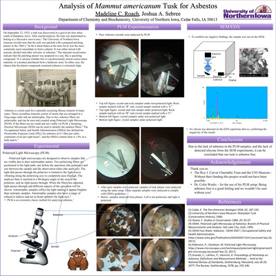
Analysis of Mammut americanum Tusk for Asbestos
Madeline C. Roach and Joshua A. Sebree
On September 23, 1933, a tusk was discovered in a gravel pit four miles south of Hampton, Iowa. After careful analysis, the tusk was determined to belong to a Mastodon americanus.1 The University of Northern Iowa museum records note that the tusk was patched with a prepared patching plaster in the 1960’s.2 In the United States at the time Alvar was the most commonly used consolidate to form a plaster. It was either mixed with acetone, alcohol and other solvents, or asbestos.3 The museum record notes indicate that the patching plaster was prepared in a can, like a spackling compound.2 It is unclear whether this is a professionally mixed conservation material, or a product purchased from a hardware store. In either case, the chance that the plaster compound contained asbestos is extremely high.
Asbestos is a term used for a naturally occurring fibrous mineral of many types. These crystalline minerals consist of atoms that are arranged in a long-range order and are antistrophic. Due to this, asbestos fibers are polarizable, and can be seen and counted using Polarized Light Microscopy (PLM). If the fibers are too small and not visible via PLM, a Scanning Electron Microscope (SEM) can be used to identify the smallest fibers.4 The Occupational Safety and Health Administration (OSHA) has defined the Permissible Exposure Limit (PEL) for asbestos at 0.1 fiber per cubic centimeter of air per eight hours5, and the OSHA content limit is 1.0% in a bulk matrix.5
-

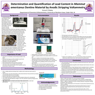
Determination and Quantification of Lead Content in Mammut americanus Dentine Material by Anodic Stripping Voltammetry
Emma R. Shipley
A 120,000-200,000 year old mastodon tusk of species Mammut americanus, recovered from Hampton, Iowa in 1933 (right) was tested for lead using a lead test from Home Depot.1 The lead test was returned positive, so Anodic Stripping Voltammetry (ASV) was undertaken to determine the concentration of lead in the dentine material of the tusk. The lead concentration is of interest because lead could affect both the health of the conservator and researchers and also inform about the diet of the mastodon. Tusk dentine material (left) functions similarly to human teeth, and studies have shown that 90% of lead in the human body concentrates in the hard tissues: skeleton and teeth.2 Lead accumulation in teeth occurs because the calcium phosphate in teeth interacts with the stable Pb isotope, causing the calcium to be permanently replaced by the lead.3 This enables teeth to be excellent records of the level of lead in the diet and environment of the organism.
-

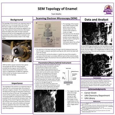
SEM Topology of Enamel
Tami Wallin
The topology of the enamel is an important topic to study how, on a microscopic level, the surface of the tusk is rough or smooth so that the correct restoration material and method are applied to the tusk. The method of study to find this information will be the SEM; which with the back scattered electrons will tell me the topography and chemical composition on the surface of the mastodon tusk fragment that will be tested, shown in the image below. SEM has better spacial resolution than optical microscopes do with the measuring tool provided by the program and the ability to clearly focus to the limits of detection in this case to a magnification of 1000 the roughness of the surface can be give measurements on how rough the topology.
